Applications research · Current
Optical diagnostics
Applying physics techniques to problems that face individual patients: testing vitamin D status at the point of need, measuring how much fat is in a donor liver before it is transplanted, and finding the receptors that mark an aggressive tumour.
Programme one
A rapid, quantitative, aptamer-based lateral flow assay for point-of-need detection of 25-hydroxyvitamin D3
Background and importance
Vitamin D (VD) deficiency is a widespread global health concern affecting more than 1 billion people and is associated with adverse skeletal, immune, metabolic, cardiovascular, respiratory and reproductive outcomes. Because VD status varies with sun exposure, season, diet, age, body composition and other factors, accessible methods for timely and repeated assessment are needed. Circulating 25-hydroxyvitamin D3 [25(OH)D3] is the primary biomarker used to assess VD status because it is stable, abundant, and reflects body VD stores.
Current methods, including immunoassays, HPLC and LC-MS/MS, provide reliable measurement but generally require centralized laboratory infrastructure, specialized instrumentation, trained personnel and sample processing. These limitations restrict rapid and decentralized testing and highlight the need for a low-cost, portable and minimally instrumented alternative.
Goal and research aims
The overarching goal is to develop a rapid, quantitative, aptamer-based lateral flow assay (LFA) for point-of-need detection of 25(OH)D3. We will:
- Aim 1: Use Capture-SELEX to identify novel 25(OH)D3-specific aptamers with target-induced structural switching and high specificity.
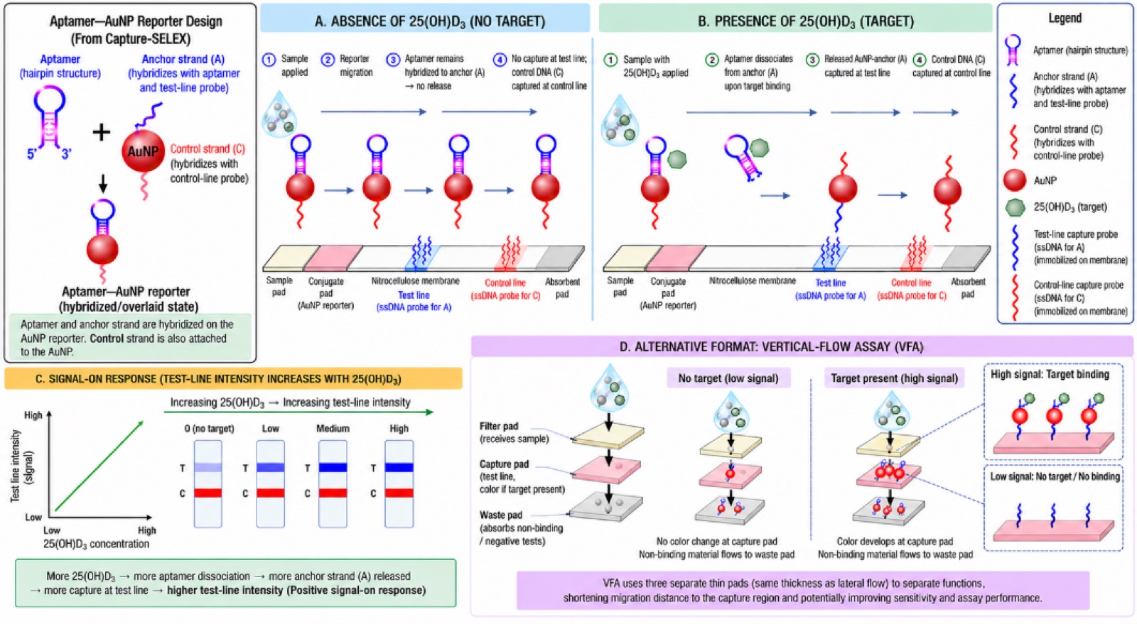
- Aim 2: Develop a target-responsive aptamer–gold nanoparticle (AuNP) reporter system that converts target binding into a measurable colorimetric signal.
- Aim 3: Integrate the reporter into a non-competitive, signal-on LFA and optimize its analytical performance.
- Aim 4: Validate the optimized assay in serum, certified reference materials, venous blood and finger-prick samples against established laboratory measurements.
Approach and expertise
Conventional antibody-based LFAs for the small molecule 25(OH)D3 require competitive formats that generate inverse signals, complicating quantitative interpretation. Aptamers offer chemically stable, reproducibly synthesized and programmable recognition elements that can potentially overcome this limitation. Rather than adapting the existing VDBA14 aptamer, which exhibits limited target-induced structural change, we will use Capture-SELEX to select aptamers specifically suited to a target-responsive sensing mechanism.
Selected aptamers will be coupled to AuNP-based reporters and incorporated into an LFA in which 25(OH)D3 binding releases the reporter, producing an increasing signal with increasing analyte concentration. The interdisciplinary team brings expertise in aptamer selection, SELEX, nanomaterials, molecular diagnostics, colorimetric biosensing and LFA development.
Expected outcomes and impact
The project will establish: (i) novel 25(OH)D3-responsive aptamers; (ii) a validated target-responsive AuNP reporter; (iii) a signal-on, non-competitive, sensitive and specific LFA over the clinically relevant working range; and (iv) proof-of-concept performance in biological samples.
The resulting platform is intended to provide rapid, low-cost, portable and minimally instrumented 25(OH)D3 testing suitable for point-of-need applications. Decentralized detection would open access to at-risk populations, including those in rural and remote communities and the global south, and the possibility of population-level monitoring for large-scale studies. Improvements in health outcomes across the over 400 processes that VD influences would enhance preventive care efforts. A 2016 estimate projected $12.5 ± $6 billion in annual economic benefit if Canadians achieved 25(OH)D levels above 100 nmol/L.
Key collaborators
Dr Malama Chisanga — Department of Chemistry, Dalhousie University.
Dr Yunyun Wu — Faculty of Dentistry, Dalhousie University.
Dr Angela Alleyne — Faculty of Science and Technology, The University of the West Indies, Cave Hill.
Dr Shane Austin — Faculty of Science and Technology, The University of the West Indies, Cave Hill.
Dr Andrea Locke — Vanderbilt University.
Programme two
Measuring liver fat in the operating theatre
A surgeon accepting a donor liver has to judge how much fat is in it. Too much fat and the organ will not function after transplant. That judgement is usually made by eye, or by a biopsy that takes time the surgery does not have.
Judging by eye is conservative — and conservative estimates discard organs that would have worked. We published the size of that effect in Transplantation in 2025: the discard rate attributable to over-estimating steatosis is large enough to matter to waiting lists.
What we built
A filter-based Raman system with lock-in amplification that reads hepatic fat content in roughly one second. Two things make it usable rather than merely accurate:
- It works in ordinary room light. Most Raman systems need darkness. Lock-in detection recovers the signal from under ambient illumination, so the theatre lights stay on.
- It costs about $18,000 rather than $65,000. Moving from a spectrometer to a filter-based design cut the instrument cost by roughly three quarters while improving reliability.
Multivariate statistical analysis does the rest — extracting fat content from the spectrum faster and more accurately than a single band ratio can. We have since shown that combining Raman with reflectance also recovers fat droplet size, which distinguishes macrovesicular from microvesicular steatosis: clinically, a different question with a different answer.
Patents were filed in 2015 and 2016. The instrument has been validated on human liver specimens across the range from minimal to severe steatosis, in work with collaborators at Leiden, the BC Cancer Agency and Vanderbilt.
Filter-based Raman analyser
785 nm excitation, filter-based detection, lock-in amplification. Built in this lab.
Key collaborators
Dr Ian Alwayn — Professor and Director, Department of Transplant Surgery, LUMC Transplant Center, Leiden, Netherlands.
Dr Haishan Zeng — Distinguished Scientist, BC Cancer Agency; Professor, University of British Columbia.
Dr Andrea Locke — Vanderbilt University.
Programme three
Nanomedicine for cancer detection
The Epidermal Growth Factor Receptor — EGFR, also called HER1 — is over-expressed in skin, lung, breast and bladder cancers. If you can see where HER1 is, you can see where the tumour is.
We build gold and silver nanoparticles bound to EGF and to anti-EGFR antibodies, and read them out by surface-enhanced Raman scattering. Two findings matter most:
- The nanoprobes show exquisite sensitivity for HER1 — they report the receptor, not the neighbourhood.
- They are not sequestered in immune organs in mouse models, which indicates low non-specific uptake. Non-specific uptake is what ruins most imaging agents before they reach a patient.
We also found that nanoparticles aggregating inside endosomes and lysosomes themselves produce surface-enhanced Raman signal — the cell assembles the hot-spot for you.
The near-term target is breast cancers that co-express HER1 and HER2. These are aggressive and poorly served by existing imaging, and a probe that reads both receptors would change how they are staged. With collaborators in Brazil we have extended the same chemistry to therapy, conjugating chlorin e6 to gold nanoparticles as a photodynamic agent for triple-negative breast cancer.
Selected publications from this area
Guo et al., “Liver discard rate due to conservative estimations of steatosis,” Transplantation 109(10), 2025.
Guo et al., “Real-time assessment of liver fat content using a filter-based Raman system, operating under ambient light through lock-in amplification,” Biomedical Optics Express 13(10), 2022.
Hewitt et al., “Accurate assessment of liver steatosis in animal models using a high throughput Raman fiber optic probe,” Analyst 140, 2015.
Related outputs
Everything this programme produced
The papers, the news and the recognition that came out of this area, each filtered to it.
The refereed papers behind this programme, filtered to optical diagnostics.
Papers, awards and the cost and clinical impact of the steatosis probe.
Press coverage of the lab’s diagnostic work.
The instruments this work is carried out on.
Funded by
Who supports this work
The agencies, foundations and partners that fund the optical diagnostics programme.
Join us
Work on this
We are recruiting a postdoctoral fellow to take the steatosis probe into a clinical study, and a research assistant for instrument build and alignment.